Understanding bone metabolism is key to diagnosing and treating various skeletal conditions. With its ability to provide detailed functional images, nuclear medicine offers unique insights into the processes underlying bone health and disease. Here is how nuclear medicine adds to our knowledge of bone metabolism:
What Is Bone Metabolism?
Bone metabolism is the process by which our bones renew themselves. It involves two main activities: bone formation and bone resorption. Osteoblasts are cells that create new bone material, while osteoclasts break down old bone tissue. This continuous cycle helps maintain strong bones and regulate calcium levels in the body.
If this balance is disturbed, it can lead to health problems such as osteoporosis, Paget’s disease, or fractures that don’t heal properly. By studying bone metabolism in detail, healthcare professionals can better understand these conditions and develop targeted treatments to manage them effectively.
What Is Nuclear Medicine?
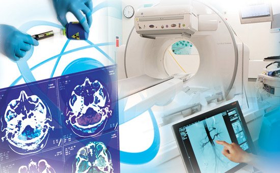
Nuclear medicine involves using small amounts of radioactive substances called tracers to examine how the body functions at a cellular level. These tracers are injected into the bloodstream and move to areas with increased biochemical activity. Special imaging devices, like gamma cameras, detect the tracers’ emissions and produce detailed images.
What Conditions Are Diagnosed?
Nuclear medicine is widely used to assess and diagnose numerous bone-related conditions. These include:
- Metastatic Bones: Cancer that has spread to the bones often triggers increased metabolic activity, making these areas glow on imaging scans.
- Osteomyelitis: This bone infection displays specific tracer patterns, helping doctors decide on the right course of antibiotics.
- Stress Fractures: Hairline cracks that are invisible on standard X-rays become evident with nuclear imaging due to increased tracer uptake.
- Arthritis Issues: Joint inflammation shows up as bright spots, providing insight into disease progression or treatment response.
Beyond detection, this technology offers clarity into how these conditions develop and affect bone tissue over time.
How Are Results Interpreted?
The interpretation of nuclear imaging depends on detecting areas where tracer uptake is either increased or decreased. This balance of activity provides clues about bone health and helps identify the cause of problems. In a patient with chronic back pain, imaging might show hot spots indicating stress fractures or degenerative issues. By turning patterns into clear data, nuclear imaging aids in accurate diagnosis and effective treatment.
Why Is It Reliable?
Nuclear medicine provides unique benefits that make it a reliable tool for studying bone metabolism. It offers real-time, functional data that other imaging methods can’t match. This means it can detect changes based on cell activity before physical changes happen. For patients, this often means earlier diagnosis and more effective treatment.
While handling radioactive substances is required, strict safety regulations and low dosage levels help minimize risks for both patients and healthcare providers. When combined with its high diagnostic accuracy, nuclear medicine is a practical and insightful way to understand bone metabolism.
Visit a Radiologist Near You
Nuclear medicine unlocks valuable insights into bone metabolism by offering a deeper look at how bones function and respond to change. With its ability to detect abnormalities early and explain complex metabolic patterns, this technology transforms patient care. By continuing to refine nuclear imaging techniques, we advance our understanding of bone health and enable more effective solutions for conditions affecting the skeletal system. Find a radiologist near you to explore body imaging options.